Diverse participation in clinical trials can advance medical research and improve community health...
Read More
Most people associate ultrasounds with getting a first peek at a baby on the way, but ultrasounds can also be used to diagnose or monitor conditions in many parts of the body, including the liver, stomach, pancreas, kidneys, breasts, pelvic area and musculoskeletal system. Our technologists are here for your ultrasound exams—close to home at all our South Jersey locations, including our outpatient AMI at Inspira imaging facilities.
Ultrasounds use high-frequency sound waves to generate images of soft tissue—something that traditional X-ray machines cannot do well. Ultrasounds are safe, painless and don’t use radiation. Technologists can perform ultrasounds at many of our locations, including our outpatient AMI at Inspira imaging locations.
During an ultrasound exam, high-frequency sound waves—far above the range of human hearing—are emitted into your body through a handheld device called a transducer. These sound waves get reflected back from your internal organs and are collected by the transducer. A computer records and interprets the resulting echoes and then generates an image of the area being examined. Most ultrasounds are conducted outside the body, but sometimes the transducer is inserted into a natural body opening, such as the esophagus, rectum or vagina, to examine internal organs.


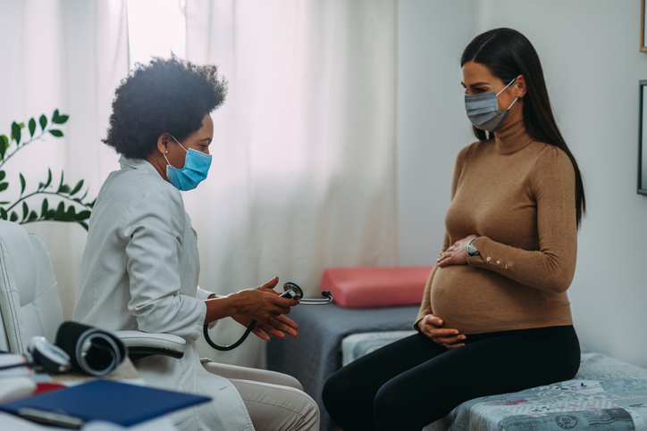
Abdominal ultrasounds are used to examine organs such as the liver, stomach and pancreas. Abdominal ultrasounds are also commonly used to monitor a baby's progress during pregnancy.

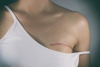
A breast ultrasound allows physicians to establish a 360-degree view of breast tissue.

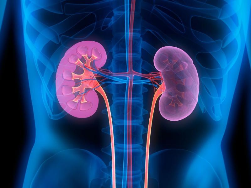
Ultrasounds of your kidneys can identify kidney stones, cysts and tumors.

Pelvic ultrasounds can examine the uterus, cervix, vagina, fallopian tubes and ovaries.

MSK Ultrasounds produce pictures of muscles, tendons, ligaments, nerves and joints throughout the body. They are used to help diagnose sprains, strains, tears, trapped nerves, arthritis and other musculoskeletal conditions.
Ultrasounds normally take just 20-30 minutes and are painless. Before your ultrasound, you may be asked to remove jewelry and clothing or change into a gown. You’ll lay on an examination table and a technologist, called a sonographer, will apply a water-based gel to the area of your body that’s being examined. This gel helps prevent air pockets, which can block sound waves.
The sonographer will place the transducer on your body and move it around to capture images. If needed, the transducer may be attached to a probe that will be inserted into your body through a natural opening, such as the esophagus to examine your heart, rectum to examine your prostate or vagina to examine your uterus and ovaries.
Ultrasounds are generally non-invasive, safe and painless. Unlike other imaging techniques, ultrasounds don’t use radiation or contrast dye.
X-rays and CT scans can provide physicians with fairly detailed images of bones and abnormalities like tumors, but aren’t very good at generating images of soft tissue. Ultrasounds, on the other hand, can show abnormalities in soft tissue and muscles and are safe for pregnant people.
Our state-of-the-art imaging technology and specialty trained, board certified radiologists can be found at all nine locations throughout South Jersey.

Diverse participation in clinical trials can advance medical research and improve community health...
Read More
Atlantic Medical Imaging (AMI), Inspira Health and Regional Diagnostic Imaging (RDI) are proud to...
Read More
If you have had lymph node surgery, especially as a treatment for breast cancer, you may be at risk...
Read More
The material set forth in this site in no way seeks to diagnose or treat illness or to serve as a substitute for professional medical care. Please speak with your health care provider if you have a health concern or if you are considering adopting any exercise program or dietary guidelines. For permission to reprint any portion of this website or to be removed from a notification list, please contact us at (856) 537-6772